Skip to main menu Skip to main content Skip to footer

Glaucoma

Happy older couple

Glaucoma

Glaucoma is a leading cause of blindness and visual impairment in the United States. Glaucoma can affect patients of all ages. Known as the “sneak thief of sight,” many people affected with glaucoma do not experience any symptoms and may not be aware that they have the disease until they have lost a significant amount of vision. With early detection and treatment, eyes can be protected against the serious loss of vision or blindness.

Glaucoma is a spectrum of disease diseases that caused optic nerve damage, resulting in visual loss and potentially blindness.

Some of the diseases or underlying conditions that can contribute to the development of glaucoma, include the following:

  • Increased pressure within the eye
  • Injury to the eye
  • Inflammatory conditions of the eye
  • High risk medications

Types of Glaucoma

There are several types of glaucoma.

Primary open-angle glaucoma, or POAG, is the most common type of glaucoma. The fluid in the eye drains too slowly through the network of tiny drainage channels, known as the trabecular meshwork. The pressure in the eye increases as the fluid in the eye continues to build. Open angle glaucoma is most often treated with topical medication (eyedrops). Interocular injection of medications and Laser Treatments are also available for those patients who are not able to tolerate topical medications, or in addition to topical medications for more severe cases. There are also surgical options both at the time of cataract surgery or as standalone procedures.

Angle closure glaucoma occurs when the drainage apparatus, known as the trabecular meshwork, becomes physically blocked by structures within the eye. This causes a sudden significant rise in the intraocular pressure. This is a painful condition and requires immediate medical attention and treatment.

Low tension, glaucoma, or LTG, is a form of open angle glaucoma.

Low tension glaucoma occurs when the optic nerve is damaged despite intraocular pressures in the “normal range”.

Secondary glaucoma develops as a complication from other medical condition, such as: eye injury, uveitis, underlying ocular disease, previous ocular surgery, diabetes, cataract, ocular tumor.

Pigmentary glaucoma occurs when pigment from the iris blocks the trabecula, resulting in the slower drainage of fluid.

Pseudoexfoliation glaucoma (PXG) is a secondary open-angle glaucoma that arises from pseudoexfoliation syndrome (PXF)—a systemic condition characterized by the abnormal accumulation of fibrillar extracellular material on the lens, iris, and other ocular structures. This material progressively clogs the trabecular meshwork, leading to increased intraocular pressure (IOP) and subsequent optic nerve damage.

PXG is often more aggressive than primary open-angle glaucoma, with higher IOP fluctuations and an increased risk of surgical complications due to weakened zonular fibers, which can lead to lens instability during cataract surgery. Careful preoperative planning and monitoring are crucial for managing this condition effectively.

Congenital glaucoma affects children who have a defect in the angle of the eye that causes the fluid to drain slowly from the eye.

Risk Factors for Glaucoma

There are several factors that contribute to the risk of developing glaucoma. They include some of the following:

  • Age
  • Ethnicity
  • Family history of glaucoma
  • Thin corneas
  • Elevated eye pressure
  • Diabetes
  • Low blood pressure
  • Medication
  • Eye injury
  • Underlying ocular condition
  • Ocular medications
  • Systemic medications
  • Myopia
  • Hyperopia

Symptoms of Glaucoma

Glaucoma symptoms depend on the type of glaucoma and how advanced it is. In the early stages, most types have no noticeable symptoms, which is why regular eye exams are critical.
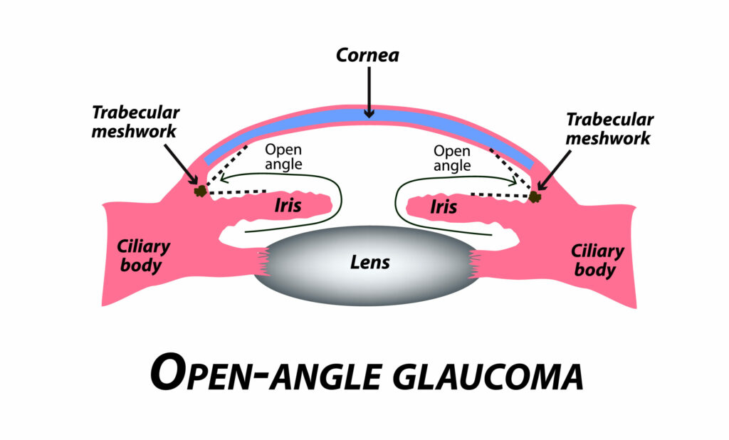
Open-Angle Glaucoma
(Most Common Type)

  • Gradual loss of peripheral vision (“tunnel vision”)
  • Blank spots in vision that gradually enlarge
  • Difficulty adjusting to dim lighting
  • Rainbow halos around lights in later stages
  • No pain or early symptoms, making it a “silent thief of sight”

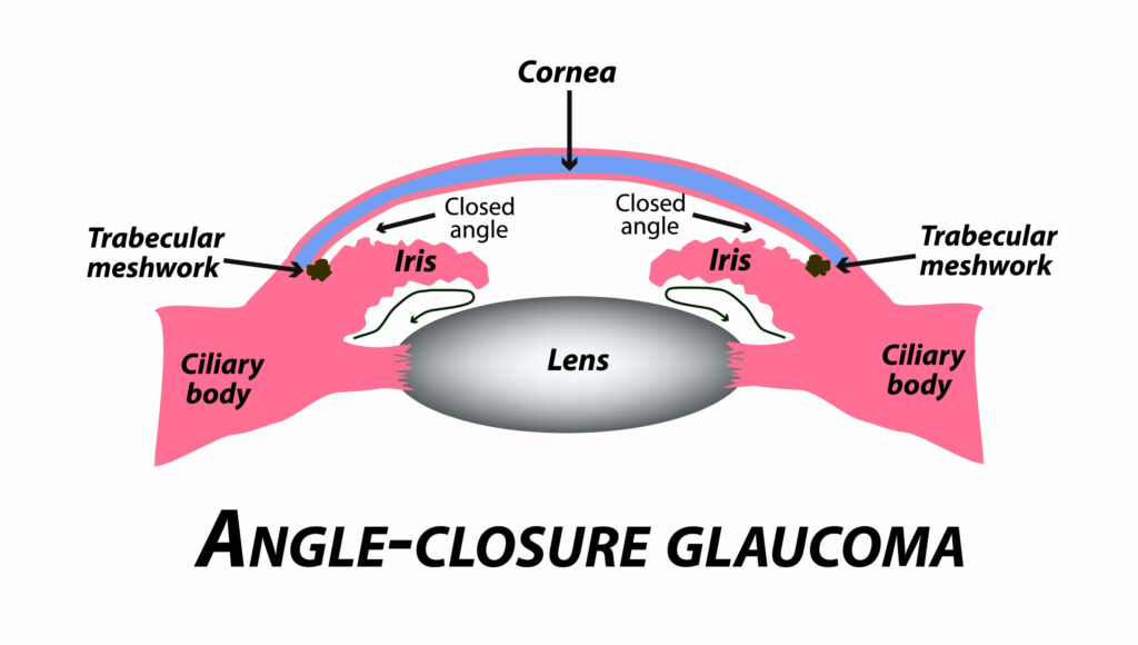
Angle-Closure Glaucoma
(Less Common, More Sudden & Severe)

  • Severe eye pain
  • Red eyes
  • Headache, nausea, and vomiting
  • Sudden blurry vision
  • Rainbow-colored halos around lights
  • Rapid vision loss (medical emergency!)
  • Tonometry: Measures intraocular pressure (IOP)
  • Gonioscopy: Examines the drainage angle of the eye
  • Visual Field Test (Perimetry): Detects peripheral vision loss
  • Ophthalmoscopy & Retinal Evaluation: Assesses the optic nerve for damage
  • Pachymetry: Measures corneal thickness (important for accurate IOP readings)
  • OCT of optic nerve and anterior segment

Treatment of Glaucoma

Although there is no cure for glaucoma, treatment can slow or stop progression and preserve vision. Early diagnosis is vital to prevent object nerve damage.

Medications

Prescription eye drops or oral medications help:

  • Reduce fluid production inside the eye
  • Improve fluid drainage to lower intraocular pressure. Side effects may include redness, irritation, stinging, or blurry vision. Consistent use is essential to maintain eye pressure control.

Laser Surgery for Glaucoma

Laser procedures improve fluid drainage or reduce fluid production. Options include:

  • Stimulates the trabecular meshwork to enhance drainage
  • Minimally invasive with little to no downtime
  • Can be repeated if needed
  • Uses thermal laser burns to improve drainage
  • Older method, less commonly used today due to scarring risks
  • Creates a small hole in the peripheral iris to prevent fluid buildup
  • Reduces risk of sudden pressure spikes and angle closure
  • Non-damaging laser pulses stimulate fluid drainage
  • Fewer side effects than traditional laser treatments
  • Targets the ciliary body to reduce fluid production
  • Micropulse CPC (MP-CPC) offers a gentler, repeatable option

Surgical Treatment for Glaucoma

When medications and laser treatments are insufficient, surgical intervention can help lower intraocular pressure and prevent further optic nerve damage. The surgical options offered at our practice include:

Minimally Invasive Glaucoma Surgery (MIGS)

MIGS procedures provide effective pressure reduction with less risk and a faster recovery compared to traditional glaucoma surgeries. We offer:

  • iStent (Glaukos): A tiny implant placed in the trabecular meshwork to improve fluid drainage.
  • Kahook Dual Blade Goniotomy (New World Medical): A bladeless procedure that removes diseased trabecular meshwork, improving fluid outflow.

MIGS procedures are often combined with cataract surgery to enhance results.

Trabeculectomy (With or Without EX-PRESS® Shunt)

Trabeculectomy is a gold-standard glaucoma surgery that creates a new drainage channel for fluid to exit the eye.

  • The EX-PRESS® Shunt (Alcon) can be used to enhance flow control, reducing complications compared to traditional trabeculectomy.

Tube Shunt Surgery (Ahmed Valve and Other Devices)

For moderate-to-severe glaucoma, a tube shunt can be implanted to divert fluid and lower pressure.

  • Ahmed Glaucoma Valve: A flow-regulating drainage implant that prevents dangerously low IOP levels.
  • Other tube shunts may be used depending on the patient’s condition and needs.

Each surgical option is tailored to the severity of glaucoma, patient anatomy, and long-term goals.

Injectables

Durysta®: A Tiny Implant That Helps Control Eye Pressure

If you’re living with open-angle glaucoma or ocular hypertension, managing eye pressure daily can feel like a constant task. Durysta® offers a breakthrough solution: it’s the first and only FDA-approved dissolvable implant that delivers glaucoma medication directly inside the eye—no need for daily drops.

What Is Durysta®?

Durysta is a prescription drug delivery system that slowly releases medication (bimatoprost) over time to lower intraocular pressure (IOP) in patients with open-angle glaucoma or high eye pressure. It’s a tiny, biodegradable implant—smaller than the “I” in the word LIBERTY on a dime! Once placed inside the eye, it gradually dissolves on its own, delivering precise dosing automatically for several months.

Note: Durysta is currently approved as a one-time treatment per eye.

How the Procedure Works

The Durysta implant is placed during a quick, in-office procedure by your eye doctor. Using a specialized applicator, we insert the implant directly into the front chamber of your eye. There are no incisions or stitches, and most patients find the experience similar to a standard eye injection.

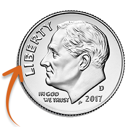
*Dime enlarged to show detail.

Once in place, the implant begins releasing medication to reduce pressure inside the eye. The entire process takes just a few minutes.

How Long Does Durysta Last?

In clinical studies, Durysta lowered eye pressure for up to 15 weeks with just one implant. While results can vary slightly from patient to patient, many people enjoy several months of consistent IOP control from a single treatment.

This makes Durysta an especially valuable option for patients who:

Benefits of Durysta

Are There Side Effects?

As with any eye procedure, there are some potential side effects. In clinical trials, the most common side effect was eye redness. Others included:

We’ll review your full medical and ocular history to determine if Durysta is a safe and effective option for you.

Is Durysta Right for You?

Durysta isn’t for everyone, but it may be a great choice if you’re looking for long-term pressure control without the hassle of daily drops. If you’ve been diagnosed with open-angle glaucoma or ocular hypertension, we’ll discuss whether you’re a good candidate during your consultation.

Let’s talk about the right treatment plan for your eyes—and your lifestyle.